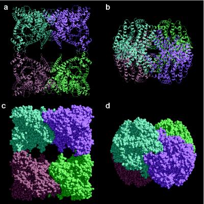
Figure 1.
Overall views of PEPC from E. coli. (a) Ribbon diagram of the homotetrameric PEPC, in which the four identical subunits colored in blue, lavender, green, and orange are related by the crystallographic twofold axes running vertically and horizontally on the page and perpendicularly to the page. (b) Ribbon diagram of tetrameric PEPC as in a but rotated 90° around a horizontal twofold axis. (c and d) Corey–Pauling–Koltun models of PEPC shown in the same orientations as a and b, respectively. The figures were produced with molscript (36) and raster3d (37).

