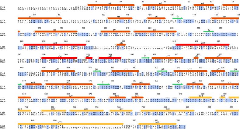
Figure 4.
Amino acid sequence alignment of E. coli and maize (C4) PEPC. The secondary structural features and residue numbers for E. coli PEPC, based on the structure reported herein, are indicated above its sequence. Residues that are identical in the two sequences are boxed in gray. Secondary structural elements of PEPC are indicated by cylinders (α-helices) or arrows (β-strands), and the missing loop from Lys-702 to Gly-708 is shown as dots. The α-helices are labeled α1–α40, and β-strands are numbered β1–β8. Color coding corresponds to that in Fig. 3. Secondary structural elements were determined with the dssp algorithm within the rasmol program (38). The figure was prepared with the program alscript (39). The specific site of phosphorylation of the maize enzyme is Ser-15 (3, 28).

