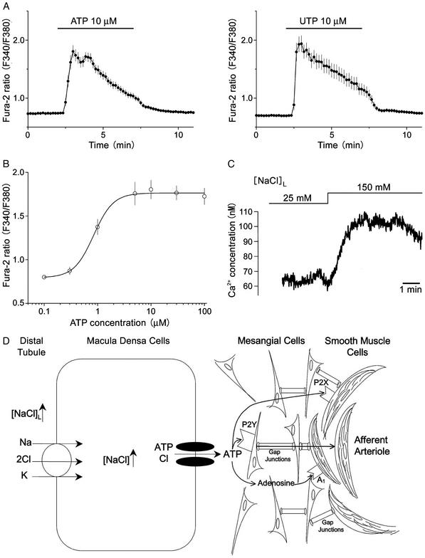
Figure 4.
The effects of ATP on cultured mesangial cells. (A) Effects of ATP and UTP on the cytosolic free Ca2+ concentration in single mesangial cells. Each symbol represents the mean values of 47 (for ATP) or 33 (for UTP) experiments, and each bar represents SEM. (B) The concentration-response curve for ATP-induced Ca2+ elevations in mesangial cells. Each symbol represents the mean value of 26–47 experiments, and each bar represents SEM. A curve represents sigmoidal fit with an EC50 value given in the text and with a Hill coefficient of 2.4. The ratio values of basal level and of peak response to 10 μM ATP correspond to the free Ca2+ concentrations of 80.2 ± 4.2 nM (n = 47) and 336.5 ± 25.5 nM (n = 47), respectively. (C) Effect of an increase in [NaCl]L from 25 to 150 mM on the cytosolic free Ca2+ concentration in a single mesangial cell that had been positioned and held with a holding pipette at the basolateral membrane of the macula densa, as shown for PC12 cells in Fig. 3C. The mean Ca2+ concentration increased from 64.7 ± 6.2 to 90.7 ± 9.7 nM (n = 5) in response to an increase in [NaCl]L from 25 to 150 mM. (D) Scheme depicting [NaCl]L-sensitive ATP release from macula densa cells via maxi anion channels at the basolateral membrane, and possible roles of released ATP in mediating signal transduction from macula densa cells to mesangial cells via P2Y receptors and to afferent arteriolar smooth muscle cells via P2X and/or A1 receptors. Signal transduction from mesangial cells to afferent arteriolar smooth muscle cells may take place via gap junctions.

