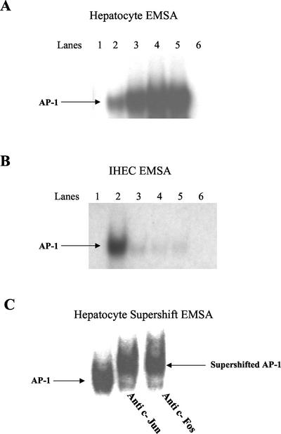
Figure 4.
EMSA analysis showing changes in DNA binding activity of AP-1 in the nuclear extracts of hepatocytes (A) and IHECs (B) stimulated via CD40 or TNF-α (positive control). Aliquots of nuclear extracts (20 μg of protein) from cultured and stimulated primary human hepatocytes and IHECs were subjected to EMSA analysis with 32P-labeled oligonucleotide probe consensus sequence to AP-1. In hepatocytes, increased AP-1 binding was detected in response to CD40 at 2 and 24 h (A), whereas in IHECs little AP-1 activation was detected in response to CD40 at either 2 or 24 h (B). (A) Hepatocyte AP-1: lane 1, negative control (labeled probe alone); lane 2, unstimulated control; lane 3, 2-h TNF-α stimulation; lane 4, 2-h CD40 stimulation; lane 5, 24-h CD40 stimulation; lane 6, cold competition with 100× excess of unlabeled probe. (B) IHEC AP-1: lane 1, negative control (labeled probe alone); lane 2, 2-h TNF-α stimulation; lane 3, unstimulated control; lane 4, 2-h CD40 stimulation; lane 5, 24-h CD40 stimulation; and lane 6, cold competition with 100× excess of unlabeled probe. (C) Supershift EMSA analysis showing the presence of AP-1 components c-Jun and c-Fos in nuclear extracts of hepatocytes stimulated for 24 h with CD40. Aliquots of nuclear extracts (20 μg of protein) from cultured and stimulated primary human hepatocytes were subjected to supershift EMSA analysis containing 2 μg of antibodies against c-Jun or c-Fos components of AP-1 and 32P-labeled oligonucleotide probe consensus sequence of AP-1.

