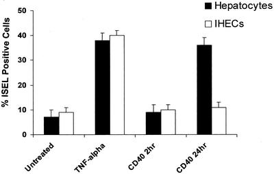
Figure 8.
Levels of apoptosis in primary cultures of hepatocytes and IHECs stimulated via CD40 or TNF-α. Histogram shows the percentage of ISEL-positive apoptotic hepatocytes or IHECs after CD40 or TNF-α stimulation. Black bars show results for hepatocytes and the white bars show results for IHECs (n = 5 for each cell type). Although TNF-α triggered similar levels of increased apoptosis in both cell types, CD40 activation only caused an increase in apoptosis in hepatocytes.

