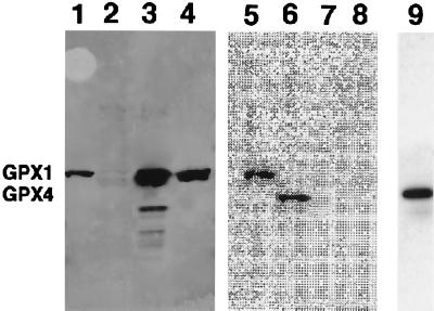
Figure 3.
Immunoblotting and imaging of GPX1 and PHGPX. Lanes 1 and 5, partially purified 75Se-labeled human T-cell GPX1; lanes 2, 6, and 9, partially purified 75Se-labeled human T-cell PHGPX; lanes 3 and 7, rat liver GPX1; lanes 4 and 8, bovine GPX1. Lanes 1–4, immunoblot assay with anti-GPX (GPX1) antibodies; lanes 5–8, PhosphorImaging of the immunoblot shown in lanes 1–4; lane 9, immunoblot detection with the anti-PHGPX antibodies. The locations of GPX1 and PHGPX (GPX4) are shown on the left.

