Figure 3.
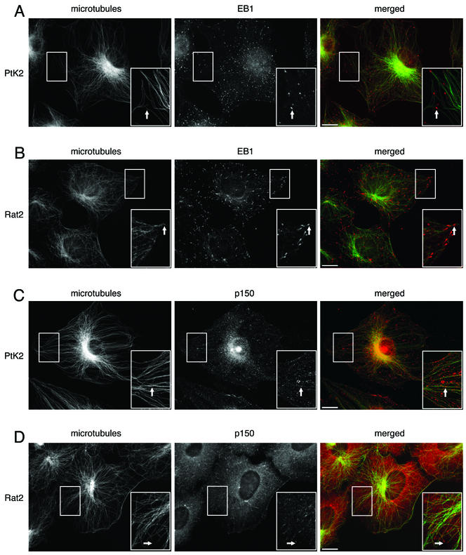
EB1 and p150Glued both localize to microtubules in epithelial cells and fibroblasts, but the pattern of localization differs. For each panel, the individual labels are shown in black and white, whereas the overlays are shown in color. The area in the box is shown magnified in the inset. Scale, 10 μm. (A) Cultured PtK2 epithelial cells labeled with antibodies to EB1 and tubulin. Arrows show EB1 (red) localized to discrete points at the plus ends of microtubules (green). (B) Cultured Rat2 fibroblasts labeled with antibodies to EB1 and tubulin. Arrows show EB1 (red) localized to comet-tails at the plus-ends of microtubules (green). (C) PtK2 cells labeled with antibodies to p150Glued and tubulin. Arrows show microtubule (green) decoration by p150Glued (red), but decoration was not limited to microtubule tips. (D) Rat2 cells labeled with antibodies to p150Glued and tubulin. Arrows show p150Glued labeling (red) was localized to comet tails at the plus-ends of microtubules (green).

