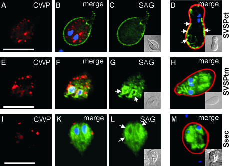
Figure 2.
Expression and transport of SVSPct, SVSPtm, and Ssec reporter proteins during Giardia encystation. CWP protein is stained with Texas red (red), SAG1 with FITC (green), and nuclei with DAPI (blue). In encysting trophozoites containing large ESVs (A), SVSPct is localized on the surface membrane (C) and the ER/nuclear envelope compartment. (B) Merged image and DAPI staining. (D) Corresponding color-merged image of a newly developed cyst <30 min after secretion of the cyst wall material. SVSPct is still partially associated with the plasma membrane (arrows). (E–G) Removal of the C-terminal pentapeptide from SVSPct (SVSPtm) leads to accumulation of the reporter in internal compartments (G), presumably ER/nuclear envelope (arrows in G), with no staining of the plasma membrane. (F) Merged images and DAPI staining. ESVs develop normally (E) and cyst formation is unimpaired (H, merged images). (I–M) Further removal of the entire VSP transmembrane domain resulted in accumulation of the now soluble reporter Ssec in the ER/nuclear envelope compartment (L, ar-rows). (I) CWP signal alone. (K) Merged images and DAPI staining. (M) Fully developed cyst showing Ssec trapped in endomembrane compartments and nuclear envelope. Insets represent differential interference contrast images of the corresponding cells. Bars, 10 μm.

