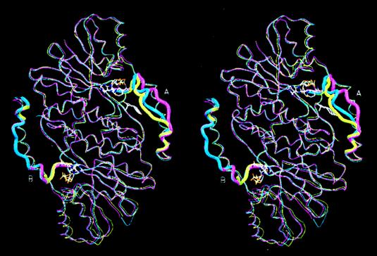
Figure 4.
Least-squares superposition of the Cα backbones of the dimers of the 17β-HSD1–equilin complex (blue), the apo-17β-HSD1 (magenta) (21)–E2-complex (yellow) (23) illustrating the differences of the substrate-entry loop structures of the A and B subunits. The substrate-entry loops (residues 186–201) are drawn in wider cross section. The loop in the A subunit of 17β-HSD1–equilin complex is in the closed conformation, whereas that in the B subunit has been modeled after the apo form. NADP+ and equilin of the 17β-HSD1–equilin complex are shown.

