Figure 1.
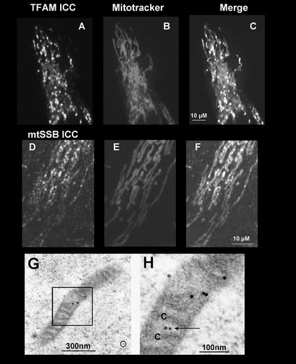
Localization of endogenous TFAM and mtSSB in intramitochondrial foci. 143B osteosarcoma cells grown on coverslips for 1–2 d were stained with Mitotracker Red (B and E). Detection of TFAM (A) and mtSSB (D) by ICC used polyclonal rabbit antibodies and a secondary fluorescein labeled antibody. (C; TFAM, F; SSB) Merged fluorescent images. (G) Immunolabeling for TFAM in a HEK293 cell mitochondrion. Seven gold particles were concentrated along a stretch of 300 nm of the mitochondrion. Very little background labeling was observed outside mitochondria (circle). (H) Blow-up of the boxed area in G, showing TFAM label (arrow) in the mitochondrial matrix near cross-sectioned cristae (C).

