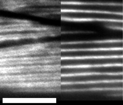
Figure 8.

Isoform-specific localization does not require the tailpiece. Adults in which relatively low levels of Δ30 expression replace endogenous MHC A [myo-3(st386); stEx150] were stained with directly labeled isoform-specific antibodies. Isoform-specific localization and overall cell structure are normal, as viewed in this portion of a muscle quadrant in which parts of three cells are visible. The left side of the micrograph shows MHC B staining along the outside edges of each A-band, and the central stripe of MHC A is shown on the right (compare with Miller et al., 1983). Bar, 5 μm.
