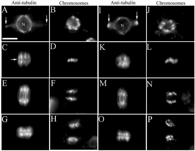
Figure 2.
Root mitosis of wild-type and mutant plants. Immunofluorescence was performed using antitubulin antibodies in dividing root cells, in both wild-type (A, C, E, and G) and homozygous mutant (I, K, M, and O) cells; the adjacent image shows the same cell stained for chromosomes (B, D, F, and H for wild-type; J, L, N, and P for mutant). In wild-type (A) and mutant cells (I), the preprophase microtubule band (arrows) and perinuclear microtubule array are present in prophase (nucleus = N). A wild-type metaphase cell (C) has a well-defined equatorial zone of microtubule clearing (arrow) and a distinct bipolar spindle with chromosomes aligned linearly along the metaphase plate (D). A mutant metaphase cell lacks an equatorial zone of microtubule clearing (K) and chromosomes do not align linearly along the metaphase plate (L); as a result there is an indistinct bipolar spindle axis. Wild-type and mutant cells have a normal anaphase spindle (E and M, respectively) and chromosomes segregate normally (F and N, respectively). Wild-type and mutant cells in telophase (G and O, respectively) show a normal microtubule-based phragmoplast and the start of chromosome decondensation (H and P, respectively). Scale bar, 10 μm.

