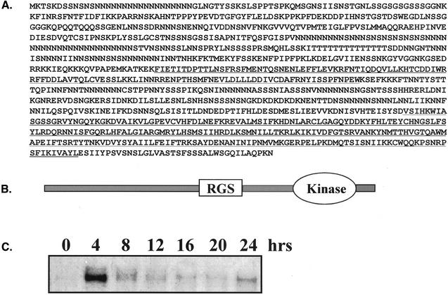
Figure 1.
Sequence analysis of Dictyostelium RCK1. (A) Deduced amino acid sequence of RCK1 from the 3990 base pairs DNA clone. The underlined region in the middle is the RGS domain. The kinase domain at the C terminus is also underlined. (B) Schematic diagram of RCK1 domain structure. (C) Northern blot showing the developmental time course of RCK1 expression. Total RNA of 8 μg/sample was resolved on a 1.0% denaturing agarose gel, blotted, and probed as described previously (Datta and Firtel, 1987). The 0-h time point is for vegetative cells.

