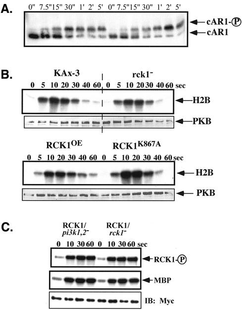
Figure 8.
(A) SDS-PAGE mobility shift assay on cAR1 phosphorylation in rck1 null cells. Cells were pulsed every 6 min for 4.5 h with 30 nM cAMP and stimulated with 100 μM of cAMP. Aliquots of cells were taken at indicated time points after adding cAMP and lysed in the same lysis buffer used for the kinase assay except that the 200 mM NaCl was omitted. SDS sample buffer was added and samples were loaded on a 12% Low-Bis SDS-PAGE gel (0.6% Bis) without boiling. Western blotting was performed using cAR1 antibodies at 1:1000 dilution. (B) PKB kinase assay in rck1 null cells and cells overexpressing wild-type and kinase-dead RCK1. Cells were pulsed with 30 nM cAMP for 4.5 h at 6-min intervals and stimulated with 100 nM cAMP. Aliquots of cells were taken at indicated time points and the PKB assay was performed as described in the Materials and Methods. For Western blotting, the same blot was probed with a rabbit polyclonal anti-PKB antibody and detected using an alkaline phosphatase-conjugated secondary antibody. (C) RCK1 activation in pi3k1/2 null cells. The RCK1 kinase activity assay was performed in pi3k1/2 null cells expressing myc-RCK1 the same way described above.

