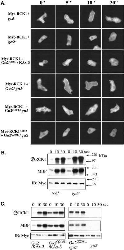
Figure 9.
(A) RCK1 translocation in various Gα backgrounds. Myc-tagged RCK1 or its mutant was either transfected alone or cotransfected with wild-type or constitutively active Gα2 (Gα2 Q208L) into various gα null cells. After establishment of positive clones, cells were pulsed for 4.5 h, stimulated with 20 μM cAMP, fixed at the time indicated, and stained for myc-RCK1 as described in MATERIALS AND METHODS. (B and C) Kinase assay of RCK1 in various Gα backgrounds as indicated. Top, autophosphorylated RCK1. Middle, RCK1-phosphorylated MBP. The immunoprecipitated RCK1 protein levels were examined by Western blot analysis on the same blot used for the kinase assay (bottom).

