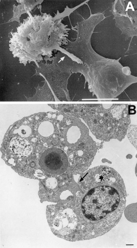
FIG. 17.
Scanning (A) and transmission (B) electron micrographs of Acanthamoeba cocultured with rat B103 neuroblastoma cells. (A) A. castellanii with extended digipodium (arrow) into the cytoplasm of the target B103 cell. (B) A. culbertsoni cocultured with B103 cells, demonstrating fingerlike projections extending into the target cell. Cells targeted by digipodia (arrow) eventually die by apoptosis or necrosis. Bars, 10 μm (A) and 1 μm (B).

