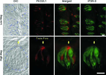
Fig. 2.
Localization of PKD2L1 protein at the taste pore. Sections of rat circumvallate taste cells were incubated with anti-PKD2L1 and anti-IP3R-3 antibodies. The dotted blue lines on the differential interference contrast (DIC) images indicate the approximate area of taste buds. Mag, magnification. (Scale bar, 20 μm.)

