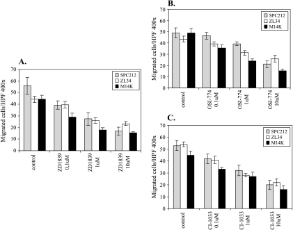
Figure 4.
(A–C) Inhibition of TGF-α-induced chemotaxis by TK inhibitors in malignant mesothelioma cells. Lower wells of Boyden chambers were filled with TGF-α (12.5 ng/ml) diluted in serum-free RPMI supplemented with 1 µg/ml BSA. The membranes were coated on both sides with 10 µg/ml of collagen type IV. Mesothelioma cells (preincubated overnight with indicated concentrations of different drugs that remained with the cells throughout the assay) were placed in the upper wells of Boyden chambers. The number of migrated cells is the mean ± SD of triplicates for each data point.

