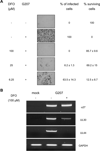
Figure 4.
Infection efficiency and cell killing of HUVEC treated with DFO at concentrations ranging from 6.25 to 100 µM. DFO was added to cell cultures 2 hours before virus infection. Infection efficiency was examined 72 hours postinfection by calculation of the percentage of lacZ-positive cells, and viable cells were counted using the trypan blue exclusion method. Each data point (mean of triplicate wells ± SD) is the percentage of cells expressing β-galactosidase or surviving cells compared with mock-infected cells in control wells, respectively (A). Effects of DFO (100 µM, added 2 hours before virus infection) on G207 replicative cycle in HUVEC. Cells were collected and cytoplasmatic RNA was extracted at 4 and 24 hours after virus infection. mRNA levels of HSV-1-specific a-gene (α27) were detected 4 hours after virus infection, whereas β-gene (UL30) and γ-gene (UL44) were measured 24 hours after virus infection. RT-PCR products were separated on a 1.5% agarose gel and visualized with ethidium bromide under ultraviolet light (B).

