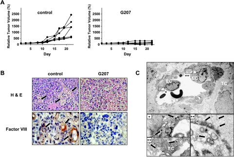
Figure 6.
Xenografts of subcutaneous KFR tumors treated with intrathecal. G207. Growth curves of control (saline-treated) and G207-treated established KFR tumors after the start of treatment (A). Examination of thin sections stained with H&E show tumor vessels filled with erythrocytes (arrows) and immunoperoxidase staining with anti -mouse Factor VIII antiserum depicts endothelial cells of tumor microvessels (arrows). Endothelial cells were not detected in tumors 8 days after G207 injection (B). Detection of G207 in endothelial cells of KFR xenografts 24 hours after G207 treatment. Enveloped viral particles are frequently enclosed in vacuoles in the cytoplasm of endothelial cells, which are shown in greater magnification in insets. Furthermore, enveloped viral particles in the cytoplasm and nucleocapsids in nucleus are shown in a tumor cell (C). EC = endothelial cell; TC = tumor cell; n = nucleus; c = cytoplasm.

