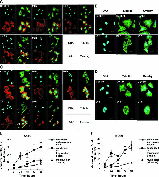
Figure 6.
Accumulation of abnormal nuclei and disturbance of microtubule and microfilament network. A549 (A and B) and H1299 (C and D) cells were exposed to 2 µM 8-Cl-Ado for 24, 48, 72, or 96 hours, respectively, and immunocytochemistry as described in Materials and Methods section. The cells were examined with a confocal microscope. Tubulin is stained with green, actin with red, and DNA with blue. The enlarged photographs of A549 (B) and H1299 (D) are presented. Cells mock-exposed for 96 and 24 hours were used as “control” in (A) and (C), and (B) and (D), respectively. (E and F) Histograms showing the percentage of abnormal nuclei relative to the total nuclei in A549 and H1299 cells, respectively. Binuclei, polymorphonuclei, and multinuclei were considered to be abnormal nuclei. Data represent mean ± SD derived from three independent experiments.

