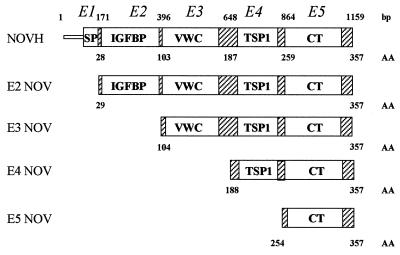
Figure 1.
Schematic representation of the NOVH full-length and truncated proteins expressed by the pGBT9 plasmids. Numbers above the structural motifs indicate the nucleotide position of NOVH exons boundaries. Numbers below the motifs indicate the amino acid boundaries of NOVH exons. VWC, Von Willebrand-type C motif; TSP1, thrombospondin type 1 motif.

