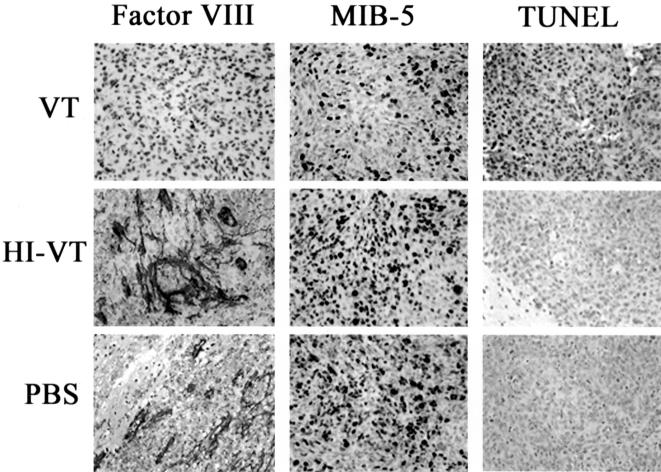
Figure 5.
Immunohistochemical analysis of VT1-associated anti-neoplastic effects in IOMM-Lee tumours. Photomicrographs (original magnification, x400) illustrates the marked reduction in MVD (Factor VIII) and the proliferation index (MIB-5) in VT1-treated animals when compared to control groups. Additionally, this figure depicts the remarkable increase in the percentage of cells undergoing apoptosis (TUNEL) in animals treated with VT1. The brown staining indicates immunoreactivity.

