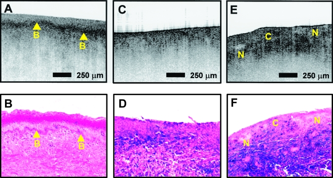
Figure 8.
Cervical disease imaged in vitro (image size: 3 mmx2 mm, resolution: 6 µm x 10 µm). (A) Carcinoma in situ is characterized by a thick, irregular epithelial layer in addition to thickening of the basement membrane (B). (C and E) Images of invasive carcinoma show a heterogeneous surface with the basement membrane no longer defined. Distinct backscattering patterns can be noted in cellular (C) and noncellular (N) regions. Images are correlated with histopathology. From Ref. [26].

