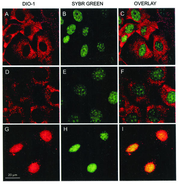
FIG. 1.
Immunolocalization of endogenous DIO-1 under apoptotic conditions. (A to C) Viable (nonapoptotic) MEF(10.1)Val5MycER cells were stained with anti-DIO-1 antibody (red); nuclei were stained with Sybr Green (green). (D to F) The same cells were induced to apoptosis by lowering the incubation temperature (32°C, 12 h) to activate p53 in the presence of 17β-estradiol (1 μM). Note the clear cytoplasmic pattern of DIO-1, although some cells have already begun the apoptotic program. (G to I) The same cell line, incubated at 39°C to inactivate p53, was 17β-estradiol treated (1 μM) and serum starved for 8 h, the time at which DIO-1 mRNA is upregulated. Note nuclear translocation of DIO-1 to the intact, nonapoptotic nuclei.

