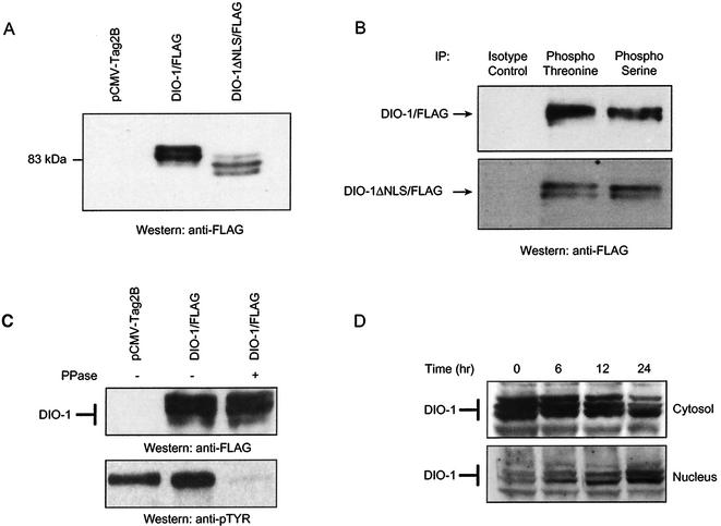
FIG. 4.
DIO-1 is present in several forms with distinct subcellular distribution. (A) Western blot of lysates from 293T cells transfected with empty vector (pCMV-Tag2B), Flag-tagged DIO-1, or Flag-tagged DIO-1ΔNLS. The membrane was blotted with anti-Flag antibody. A representative result of five independent experiments is shown. Note the lower apparent molecular size of the deletion mutant and the three-band pattern. (B) 293T cells were transiently transfected with the indicated Flag-tagged constructs. Lysates were immunoprecipitated with antiphosphothreonine, antiphosphoserine, or an irrelevant control antibody and then blotted with anti-Flag monoclonal antibody. Only the upper and middle bands were detected. (C) 293T cells were transiently transfected with Flag-tagged DIO-1. Untreated or λ-phosphatase-treated lysates were separated by SDS-PAGE and then blotted with anti-Flag monoclonal antibody. Phosphatase treatment did not cause a mobility shift (upper panel). Dephosphorylation of total cellular proteins was verified by blotting with antiphosphotyrosine antibody (lower panel). (D) FL5.12 cells deprived of IL-3 for the times indicated were separated into cytosolic and nuclear fractions (Materials and Methods). Extracts were separated by SDS-PAGE and analyzed by Western blotting with the anti-DIO-1 antibody.

