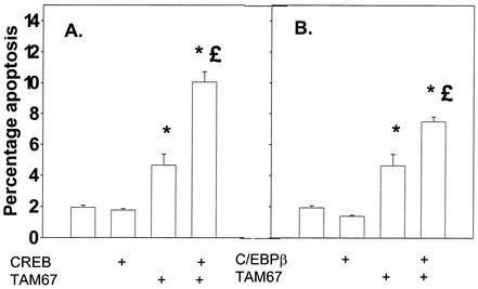
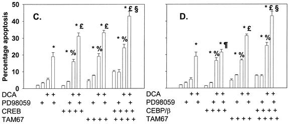
FIG. 8.
Dominant-negative c-Jun (TAM67) can interact with either dominant-negative CREB or dominant-negative C/EBPβ to enhance the apoptotic response following DCA exposure. Hepatocytes were isolated and cultured as described in “Methods.” As indicated, 4 h after plating, cells were infected either with control plasmid- and poly-l-lysine-conjugated adenovirus, with dominant-negative c-Jun (TAM67) plasmid- and poly-l-lysine-conjugated adenovirus, with dominant-negative CREB plasmid- and poly-l-lysine-conjugated adenovirus, or with dominant-negative C/EBPβ plasmid- and poly-l-lysine-conjugated adenovirus at a total combined multiplicity of infection of 500. Twenty-four hours after plating, cells were treated with either vehicle or vehicle containing the MEK1/2 inhibitor PD98059 (final concentration, 50 μM). Thirty minutes after kinase inhibitor treatment and 24 h after adenovirus infection, cells were exposed to bile acid (final concentration, 50 μM). (A and B) Expression of dominant-negative c-Jun (TAM67) enhances the basal apoptosis level that is augmented by expression of either dominant-negative CREB or dominant-negative C/EBPβ. (C and D) Expression of dominant-negative c-Jun (TAM67) enhances DCA-induced and DCA-plus-MEK1/2 inhibitor-induced apoptosis levels that are augmented in a less-than-additive fashion by expression of either dominant-negative CREB or dominant-negative C/EBPβ. *, P < 0.05, greater than vehicle-treated control; %, P < 0.05, greater than corresponding value for cells not expressing dominant-negative transcription factor; %, P > 0.05, no statistical difference between vector control cell treated with DCA and MEK1/2 inhibitor and dominant-negative transcription factor-expressing cells treated with DCA alone; £, P < 0.05, greater than vector control cell treated with DCA and MEK1/2 inhibitor; §, P < 0.05, greater than parallel value for treated cells expressing an individual dominant-negative transcription factor.


