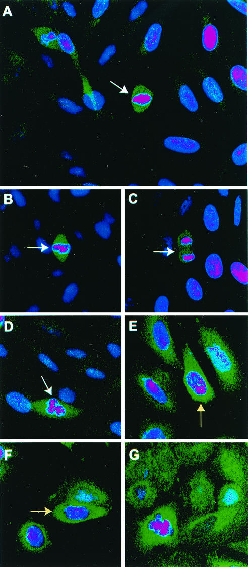
FIG. 1.
Staining of cells with SIRT2 antibody is tightly confined to cells in late G2 or M phase. Confocal microscopy was performed on Saos2 cells grown on coverslips as described in Materials and Methods. DAPI (dihydrochloride)-stained nuclei appear pink. DAPI stain is blue, but in the interests of contrast, the color was enhanced. A green fluorescent FITC-conjugated goat anti-rabbit secondary antibody was used to localize the rabbit SIRT2 peptide primary antibody. White arrows point to mitotic cells. (A) Full-field view of unsynchronized Saos2 cells. Only mitotic cells exhibit intense SIRT2 staining. (B to D) Close-up views of individual Saos2 mitotic cells. (E and F) Saos2 cells blocked at G2/M with nocodazole. The intense SIRT2 staining tightly surrounds the condensing chromatin (indicated by yellow arrows). (G) Saos2 cells blocked in metaphase with Colcemid. Again, SIRT2 is intensified around the chromatin.

