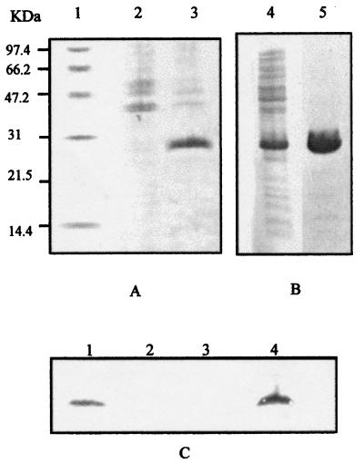
Figure 1.
Electrophoretic characterization of MsrA protein. (A) SDS/PAGE analysis of E. coli cultures grown in LB medium with added IPTG (1 mM). Proteins were visualized by Coomassie staining. Lane 1: size markers; lane 2: E. coli BL21/pET22b+; lane 3: E. coli BL21/pTMS10. (B) SDS/PAGE analysis of the purification of MsrA-(His)6. Lane 1: BL21/pTMS10 extract; lane 2, MsrA-(His)6 protein eluted from the Ni-NTA resin after treatment with imidazol. (C) Immunoblot analysis of E. chrysanthemi strains 3937 (lane 1), MEH14 (lane 2), MEH14/pLAFR3 (lane 3), MEH14/pLAMS11 (lane 4) by using anti-MsrA antibodies.

