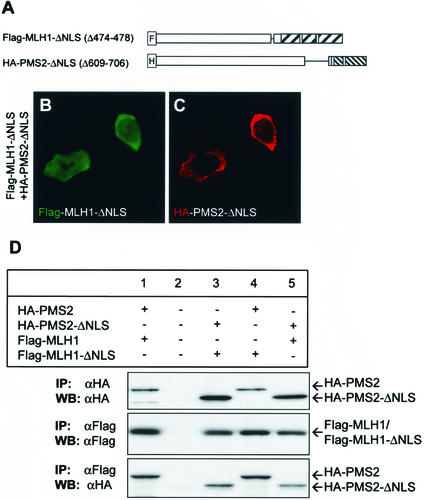
FIG. 7.
NLS of MLH1 and PMS2 are required for nuclear localization. (A) Schematic representation of NLS MLH1 and PMS2 deletion mutant constructs Flag-MLH1-ΔNLS and HA-PMS2-ΔNLS, respectively. (B and C) Intracellular localization of MLH1 (green) or PMS2 (red) mutants in HCT 116 cells cotransfected with Flag-MLH1-ΔNLS and HA-PMS2-ΔNLS. MLH1 or PMS2 was detected with anti-Flag (B) or anti-HA (C) antibodies. Images B and C represent 439 (88%) of 500 transfected cells examined and were collected and processed by using the same settings, as explained in Materials and Methods. (D) Western blot analysis of MLH1 and PMS2 immunoprecipitated from nontransfected HCT 116 cells (lane 2), cells transfected with full-length HA-PMS2 (lanes 1 and 4) and Flag-MLH1 (lane 1) or Flag-MLH1-ΔNLS (lane 4), and cells transfected with HA-PMS2-ΔNLS (lanes 3 and 5) and Flag-MLH1-ΔNLS (lane 3) or Flag-MLH1 (lane 5). Whole-cell lysates were immunoprecipitated (IP) with anti-Flag or anti-HA antibodies, as indicated. Precipitated proteins were resolved on SDS-8% PAGE and were blotted onto PVDF membranes (WB), followed by probing with anti-Flag or anti-HA antibodies, as indicated. Bands were visualized with the ECL detection kit.

