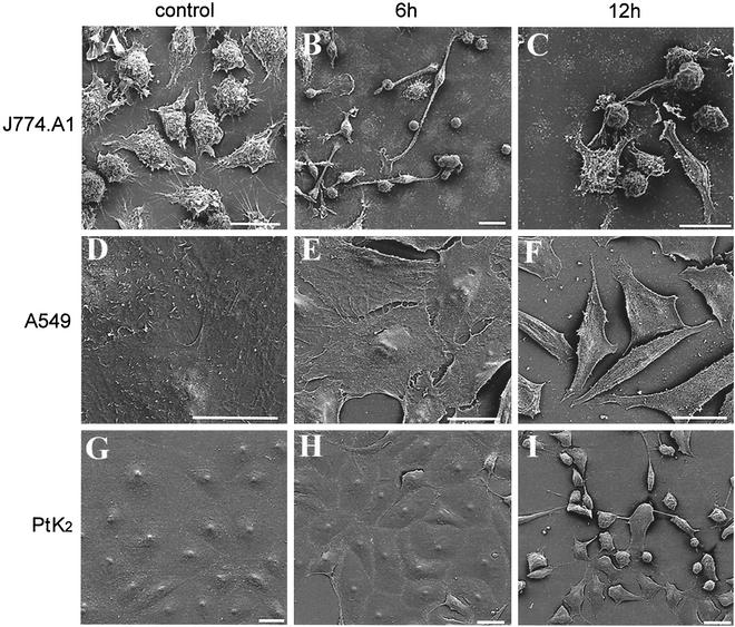
FIG. 1.
Morphological changes induced by B. pseudomallei rhamnolipid. J774.A1 (A), A549 (D), and PtK2 (G) control cells and cells exposed to B. pseudomallei rhamnolipid for 6 h (B, E, and H) and 12 h (C, F, and I) were observed by scanning electron microscopy. Rhamnolipid exposure led to a progressive rounding up and, finally, detachment in all three cell lines tested. Bar = 20 μm.

