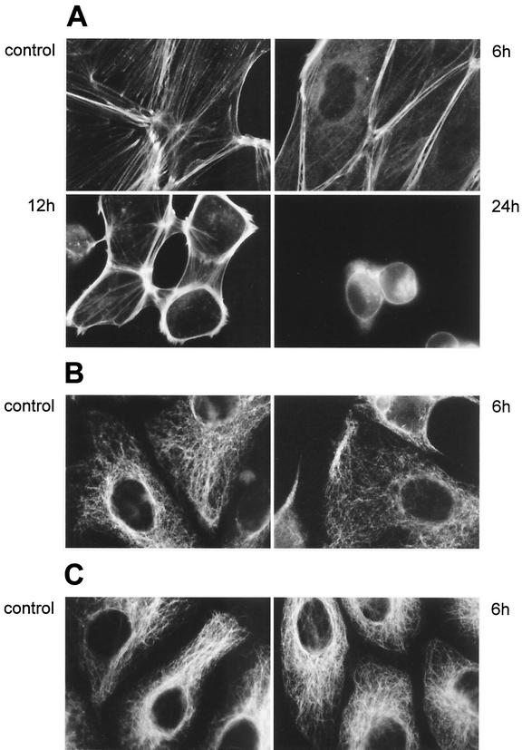
FIG. 2.
Fluorescence microscopy of the cytoskeleton in rhamnolipid-treated cells. (A) Fluorescence-labeled filamentous actins of both control PtK2 cells and cells exposed to B. pseudomallei rhamnolipid for 6, 12, and 24 h demonstrate a progressive reorganization of the actin network after intoxication. (B and C) Fluorescence-labeled intermediate (B) and microtubule (C) filaments in both control PtK2 cells and cells exposed to rhamnolipid for 6 h. Rhamnolipid exposure did not seem to induce primary morphological changes in the microtubule and intermediate-filament networks.

