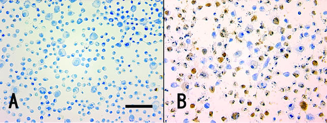
FIG. 1.
Apoptotic in situ DNA fragmentation analysis. Cytospin was carried out to attach the apoptotic cells to the glass slide, and apoptosis was monitored by the TUNEL assay. The percentage of TUNEL-positive cells (brown) was 0.60% ± 0.15% in the control (in the absence of Stx1) (A) and 63.7% ± 2.7% after exposure of the cells to Stx1 (10 ng/ml) for 4 h (B).

