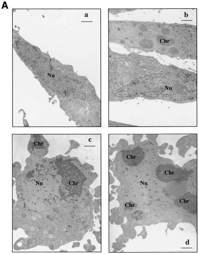
FIG. 2.
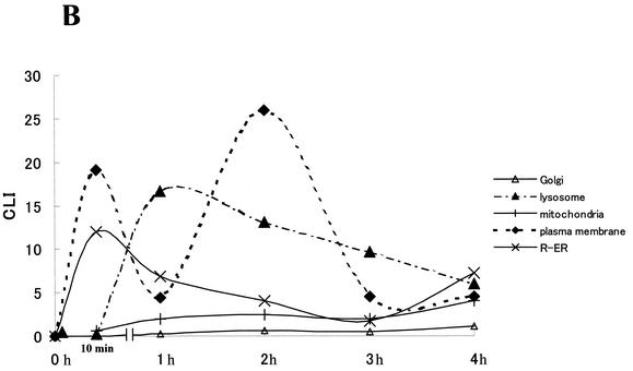
(A) Morphology of Stx2-treated HeLa cells. (a) Control. Note the fibroblastic appearance typical of untreated cells with the characteristic oblong euchromatic nucleus (Nu). (b through d) Cells treated with Stx2 (10 ng/ml) for 4 h. (b) The lower cell has a morphology similar to that of a control cell. In contrast, note that in the upper cell the chromatin (Chr) has started to aggregate in fine granular masses, yet the cell maintains its fibroblastic shape. (c) The cell has begun to round up, and the cytoplasm has started to bleb. Within the nucleus, note the fine granular chromatin concentrated in the periphery of the nucleus associated with the nuclear envelope. Also evident is a fragment of the chromatin associated with a cytoplasmic bleb in the right-hand corner of the micrograph. (d) The fine granular masses of chromatin are associated with the nuclear envelope. The cell shown is rounded, and the cytoplasm has begun to bleb. The morphology shown in panels c and d was characteristic of >50% of the cells viewed in samples 4 h after treatment with Stx2. Bars, 1 μm; final magnification, ×7,500. (B) Stx2 trafficking in HeLa cells analyzed by immunogold electron microscopy. Stx2A distribution was quantified in cytoplasmic compartments such as the Golgi apparatus, lysosome, mitochondria, plasma membrane, and R-ER.


