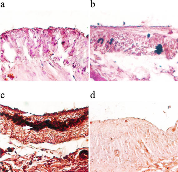
FIG. 5.
Representative slides showing immunostaining of fibrinogen in cecal tissue 24 h after surgery of sham controls (a), CLP plus irrelevant antibody (b), and CLP plus IFN-γ antibody (c) and in the section from a CLP not incubated with primary antibody against fibrinogen but instead with irrelevant antibody (d). Arrows point to areas of dark brown staining showing fibrinogen staining. The photos show that there was increased fibrin deposition in cecal tissue 24 h after CLP compared to that in sham controls. IFN-γ antibody administration further increased fibrin deposition in cecal tissue. The staining was specific for fibrin, as there was virtually no positive staining in slides that were not incubated with primary antibody.

