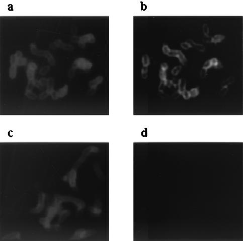
FIG. 6.
Immunofluorescence microscopy of SUNY 465 and the Aae mutant. (a and b) Wild-type cells treated with anti-SUNY 465 antibody (a) and anti-rAaePD antibody (b). (c and d) Aae mutant treated with anti-SUNY 465 antibody (c) and anti-rAaePD antibody (d). Primary antibodies were used at a 1:10,000 dilution, secondary antibodies were used at a 1:100 dilution, and blue fluorophore-conjugated antibody was used at a 1:100 dilution.

