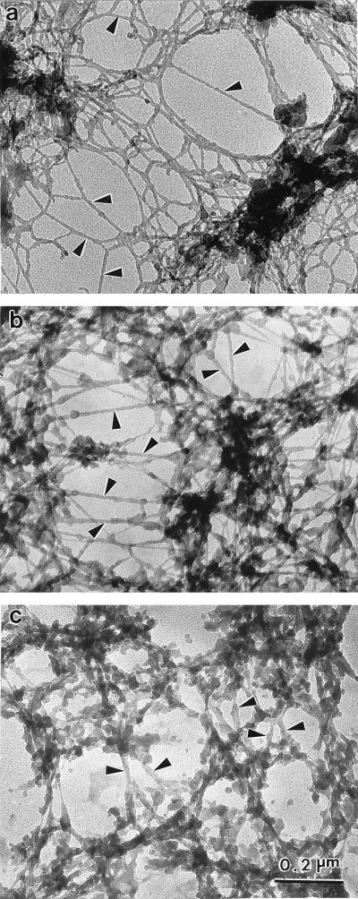
Figure 5.
Ultrastructural comparison of nuclear matrices prepared by three methods. CaSki cell nuclear matrices were prepared by three different methods and then visualized by resinless section EM. Higher magnification views of the nuclear interior are presented. (a) The high-salt nuclear matrix prepared by extracting cells with 0.5% Triton X-100 and then removing chromatin by digestion with PstI/HaeIII and sequential extractions with 0.25 M ammonium sulfate and 2 M NaCl. This procedure uncovered a network of branched filaments with an average diameter of 10 nm (arrowheads). The network of filaments connects to the inside of the nuclear lamina (not shown). (b) The amine-modified nuclear matrix was prepared in a similar way, except that the salt extractions were replaced by treatment with sulfo-NHS. The fundamental structural element revealed was a network of branched core filaments (arrowheads). More material coating the core filaments was retained. (c) The crosslink-stabilized nuclear matrix was prepared (6). After a 0.5% Triton X-100 extraction to remove soluble proteins, the structural networks were extensively crosslinked with formaldehyde. Chromatin was then removed by DNase I digestion. The more intricate structure of thicker fibers was built on an underlying network of core filaments (arrowheads). All three nuclear matrix methods reveal similar principles of nuclear construction with the nuclear matrix built on a network of branched core filaments connected to the nuclear lamina.

