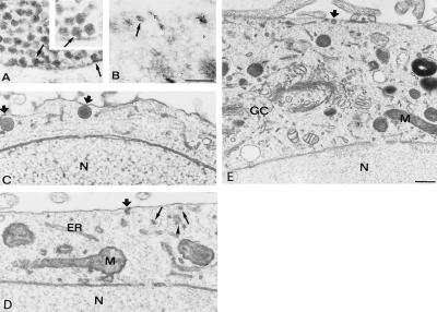
Figure 3.
Electron microscopy of a frog neuromuscular junction (A) and wild-type PC12 cells (B–D) physically fixed by qf-fd while at rest. A shows aggregation of synaptic SVs in the proximity or in direct contact [docking shown by arrows in the main panel as well as in the high magnification (Inset, ×100,000)] with the presynaptic plasma membrane. B shows a wild-type PC12 processed as described in ref. 18, immunogold-labeled for synaptophysin, a protein of SV (arrow) membranes. Additional immunolabeling decorates structures adjacent or perpendicular to the plasmalemma, most likely participating in endocytosis (13). C–D show images of wild-type PC12 cells illustrating the docking to the plasmalemma of two LVs (thick arrows in C) and of a single SV (thick arrow in D), with additional SV profiles (arrows) present in the juxtaplasmalemma layer of the cytoplasm; the arrowhead points to a cluster of apparently interconnected profiles, possibly of ER nature. E shows a vast cytoplasmic area including the section of the Golgi complex (GC) and trans-Golgi network. A docked LV is pointed by the thick arrow. N, nucleus; M, mitochondrion. [Bars = 0.3 μm (A–D).] Morphometry of data is reported in Table 1.

