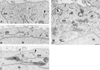
Figure 4.
Electron microscopy of defective PC12-27 cells physically fixed by qf-fq while at rest. Notice that, in all panels, the cytoplasmic layer adjacent to the plasmalemma is almost completely devoid of vesicles profiles. In contrast, profiles are numerous in the deeper layers of the cytoplasm in which other organelles (mitochondria and ER cisternae) are also visible. Profiles that might be V27 are pointed out by arrows, and those that might be ER clusters are pointed out by arrowheads. A well developed Golgi complex (GC), with its adjacent trans-Golgi network, shows a large complement of vesicles (D). [Bars = 0.3 μm (A–C).] N, nucleus; M, mitochondrion; LB, lipid body. Morphometry is reported in Table 1.

