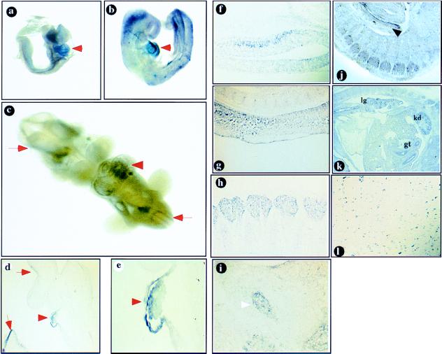
Figure 4.
(a–e) Whole-mount RNA in situ hybridization analysis of FOG-2 expression. FOG-2 is expressed in the developing mouse heart (arrowheads) in E8.5 (a), E9.5 (b, d, e), and E10.5 (c) embryos. The expression in the developing nervous system is shown by arrows (c–e). The embryo in b was sectioned to demonstrate the expression in the developing pericardium and myocardium (d, ×100, e, ×400).(f–l) In situ hybridization of FOG-2 antisense RNA probe to mouse tissue sections.(f–i) sagittal sections of an E11.5 embryo. Expression is detected in the midbrain neurons (f), spinal cord (g), dorsal root (h), and trigeminal (i, arrowhead) ganglia. Expression in the urogenital ridge is indicated by an arrow (j). (k) Sagittal section of an E15.5 embryo. Expression is detected in the developing lung (lg), kidney (kd), and gut (gt). (l) FOG-2 expression in the postnatal (P21) brainstem.

