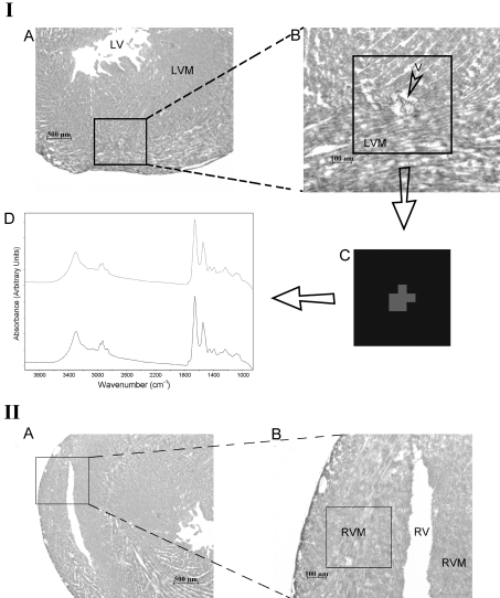
Figure 2. Light microscope images and mapped regions of the rat heart.
(IA) Light microscope image of H/E-stained thin slice taken from the left ventricle at ×25 magnification. (IB) The mapped region of the left ventricle myocardium at ×100 magnification. (IC) The image of cluster analysis in 950–1480 cm−1 region. (ID) The average spectrum arising from different clusters. (IIA) Light microscope image of H/E-stained thin slice taken from the right ventricle of rat heart at ×25 magnification. (IIB) The same image including the mapped region at ×100 magnification. Abbreviations: LV, left ventricle; LVM, left ventricle myocardium; V, vein; RV, right ventricle; RVM, right ventricle myocardium.

