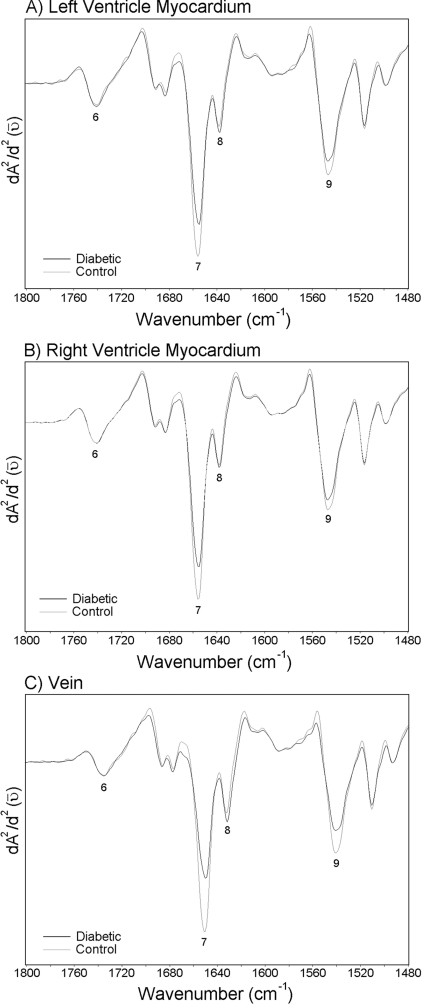
Figure 4. Average spectra belonging to the left ventricle myocardium, the right ventricle myocardium and the vein of the control and the diabetic groups in the 1480–1800 cm−1 region.
Vector normalization was done in the 950–1480 cm−1 region. Absorption maxima appear as minima in the second derivatives. (A) Second derivative average spectra belonging to the left ventricle myocardium; (B) second derivative average spectra belonging to the right ventricle myocardium; (C) second derivative average spectra belonging to the vein.

