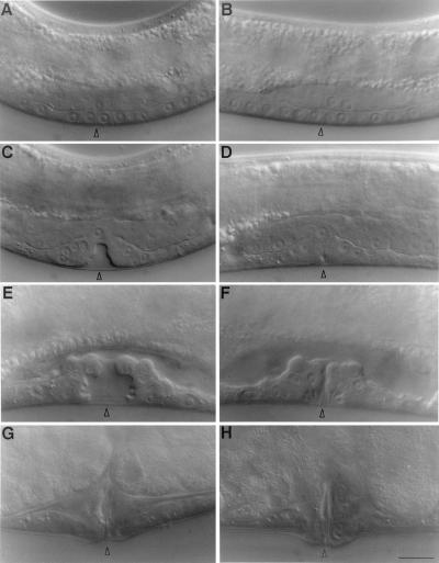
Figure 2.
The sqv mutations result in a partially collapsed vulval invagination. Nomarski micrographs of wild-type (A, C, E, and G) and sqv-3(n2841) (B, D, F, and H) vulvae before, during, and after vulval invagination. (A and B) Late L3 stage. In both wild-type and sqv mutant vulvae the vulval precursors P5.p, P6.p, and P7.p all have given rise to four cells, which lie in a plane with the surrounding epithelium, hyp7. The arrowhead points to the center of the vulva, between cells P6.pap and P6.ppa. In the wild type, the gonadal anchor cell eventually takes a position directly above and between these cells, as seen in A. The anchor cell in the sqv-3(n2841) animal in B has not yet taken this position and lies slightly posterior. The animal in B is also homozygous for unc-69(e587am). (C and D) Early L4 stage. Vulval cell division is complete. In both the wild type and the sqv mutant the appropriate cells have detached from the cuticle and left the plane of the surrounding epithelium, but the space between the vulval cells and cuticle is considerably smaller in the sqv mutant than in the wild type. Again, the arrowhead points the center of the vulva. (E and F) Mid-L4 stage. The sqv mutant invagination appears to extend less dorsally than does the wild type, suggesting that the former is partially collapsed and that the space between the vulval cells and cuticle is reduced at least in part as a consequence. (G and H) Adult. The wild-type and sqv mutant vulvae do not obviously differ in appearance, although in later adulthood, when sqv mutants become bloated with eggs, their vulvae often protrude abnormally. The bar at the lower right represents 10 μm, and the animals are oriented such that anterior is to the left, and ventral is down.

