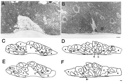
Figure 3.
The vulval invagination space is more electron dense, and the central vulval cells are more elongated in a sqv mutant than in the wild type. Electron micrographs of a wild-type (A) and a sqv-3(n2842) mutant (B) early L4 vulva in the plane at which the invagination space is largest. In each case the invagination space is the region of decreased electron density, and the cuticle is the ventral-most narrow gray band. Sections were cut so that each was parallel to the plane of the Nomarski micrographs in Fig. 2. The appropriate sqv-3(n2842) vulval cells have detached from the cuticle, but the resulting invagination space is reduced in size and more electron dense than that of the wild type. Tracings of vulval plasma and nuclear membranes and the cuticle from electron micrographs of a wild-type animal (C and E) and a sqv-3 mutant animal (D and F). Cell identifications were made on the basis of the known arrangement of vulval nuclei as judged from Nomarski DIC microscopy, and each cell is labeled with the letter name of the corresponding late L4 toroid (42). Tracings C and D are of the micrographs partly shown in A and B, and tracings E and F are of sections located roughly the same lateral distance from those traced in A and B, respectively. The central sqv-3 vulval cells are abnormally elongated, extending into and reducing the size of the invagination space. The bars at the lower right of B and F represent 1 μm.

