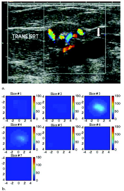
Figure 7.

US image and hemoglobin concentration maps of suspicious 1.9 × 1.1-cm lesion at 9 o’clock position in right breast of a 75-year-old woman. (a) US image demonstrates lesion with solid component adjacent to cyst (arrow). Large blood vessels were seen at periphery of lesion during color Doppler US. Biopsy result revealed intraductal papilloma, with no evidence of atypical cells or malignancy. (b) Total hemoglobin concentration map computed from absorption maps (not shown) obtained at 780 nm and 830 nm. Vertical scale presents total hemoglobin concentration in micromoles and ranges from 0 to 150 μmol/L. Maximum total hemoglobin concentration was 38.2 μmol/L, and average total hemoglobin concentration was 26.3 μmol/L, as measured within FWHM region. Total hemoglobin distribution was diffused rather than localized, as seen in cases of malignancy.
