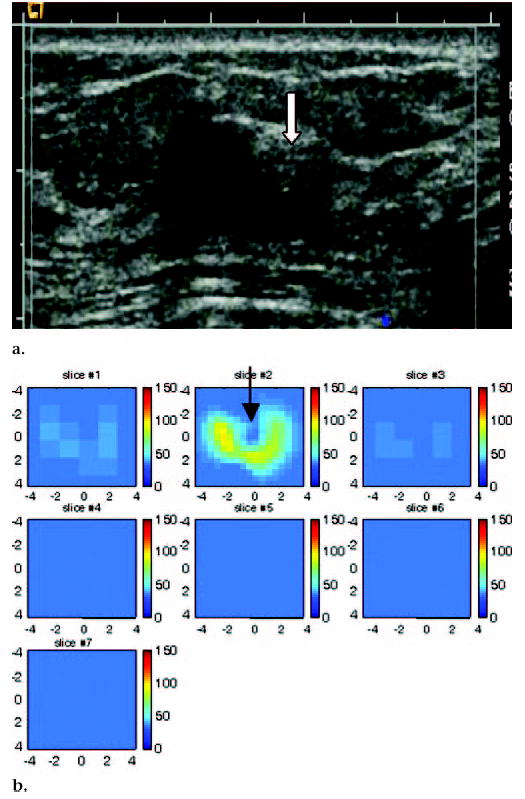
Figure 8.

US image and hemoglobin concentration maps of 1.3 × 1.0-cm lesion, simple cyst, and complex cyst located between 3 o’clock and 4 o’clock positions in left breast of 59-year-old woman. (a) US image demonstrates simple cyst with adjacent complex cyst or solid nodule (arrow). Complex cyst was aspirated completely, and no residual abnormality was noted. (b) Total hemoglobin concentration map indicates low hemoglobin concentration at central location of cyst (arrow) close to origin in section 2 (slice #2). Light absorption maps (not shown) indicated low light absorption. Incomplete ring with higher light absorption and high hemoglobin concentration was observed surrounding cyst. Although measured total hemoglobin concentration was highest in complex cyst owing to higher absorption ring, distribution was obviously different from malignant cases (maximum total hemoglobin concentration, 94 μmol/L; average total hemoglobin concentration, 62.8 μmol/L, as measured within FWHM region). Cysts, in general, have low light absorption (and therefore low hemoglobin concentration) owing to low water absorption in wavelength range studied. The higher absorption ring was likely caused by the cyst wall. This example suggests that both hemoglobin distribution and threshold level must be evaluated for accurate diagnosis of suspicious lesions.
