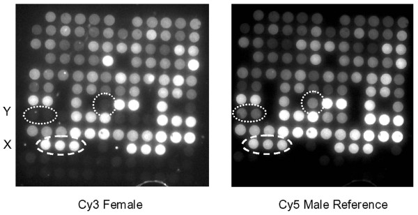
Figure 2.

Array MAPH images. Typical image-captures of a single array from a PMP22 PamChip after co-hybridization with a Cy3 labelled female patient sample (left) and the Cy5 labelled male reference sample (right). Spots containing oligonucleotides complementary to the Y and X chromosomes are indicated. The 12 × 12 arrays shown are composed of duplicate spots of 72 oligonucleotides (in 6 columns and 12 rows), such that the left and right halves of the array are identical. The oligonucleotides occupying the bottom two rows are control sequences not complementary to the probes applied.
