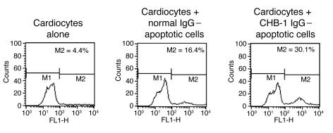
Figure 5. Use of annexin V to assess the persistence of opsonized and nonopsonized apoptotic cardiocytes after incubation with proliferating cardiocytes (5 hours).
Apoptotic cardiocytes were treated with normal control IgG (nonopsonized) or with IgG from an anti-SSA/Ro– and -SSB/La–positive CHB mother (CHB-1; opsonized). Monolayer proliferating cardiocytes were incubated alone, with opsonized apoptotic cells (CHB-1 IgG–apoptotic cells), and with nonopsonized apoptotic cells (normal IgG–apoptotic cells). After 5 hours, cells were scraped from the culture dish and stained using annexin V. M1 and M2 indicate gates of FACS analysis.

