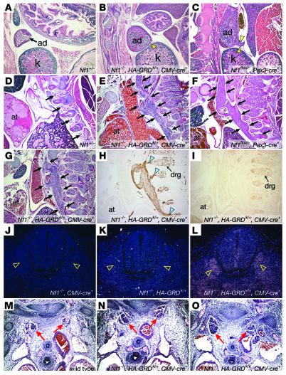
Figure 4. HA-GRD does not rescue abnormal neural crest–derived tissues lacking Nf1.
(A–C) Adrenal medullary enlargement in HA-GRD rescued newborns. Sagittal sections of adrenal glands (ad); Nf1–/–, HA-GRDk/+, CMV-cre+ (B) and the Nf1 neural crest–specific knockout, Nf1flox/–, Pax3-cre+ (C) had overgrown adrenal medullae compared with wild-type (A) with cortical effacement and tumor-like medullary protrusion (arrowheads). k, kidney. (D–I) Enlarged peripheral ganglia in HA-GRD rescued newborns. Sagittal sections of Nf1+/– (D); Nf1–/–, HA-GRDk/+, CMV-cre+ (E, G–I); and Nf1flox/–, Pax3-cre+ (F). at, cardiac atrium. Peripheral ganglia (arrows) were massively enlarged in Nf1–/–, HA-GRDk/+, CMV-cre+ newborns (E and G) compared with an Nf1+/– (D) littermate and similar the Nf1 neural crest–specific knockout (F). (H) Positive staining for neurofilament, with connections (arrowheads) to dorsal root ganglia (drg). (I) Negative staining for the glial marker S-100. (J–L) HA-GRD is expressed with CMV-cre. In situ hybridization of cross-sections of Nf1–/– E12.5 embryos with CMV-cre transgene (J), HA-GRD knock-in (K), or both (L) show expression of HA-GRD only with both. Dorsal root ganglia (arrowheads) had notably robust expression. (M–O) HA-GRD dose did not alter abnormalities of neural crest–derived tissue in Nf1–/– embryos. Cross-sections of wild-type (M); Nf1–/–, HA-GRDk/–, CMV-cre+ (N); and Nf1–/–, HA-GRDk/k, CMV-cre+ (O) E12.5 embryos highlighting sympathetic ganglia (red arrows), enlarged in both Nf1–/– genotypes and no smaller with 2 copies of HA-GRD (O) than with 1 copy (N). Magnification, ×4 (A–I); ×10 (J–O).

