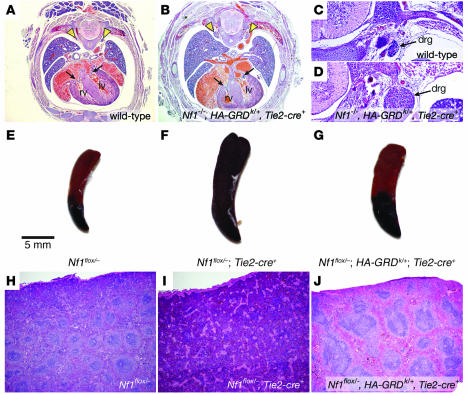
Figure 5. Tie2-cre –mediated expression of HA-GRD rescues cardiac development and hematologic abnormalities.
(A and B) Cardiac rescue with endothelial expression of HA-GRD. Representative low-power photomicrographs (magnification, ×4) of E17.5 wild-type (A) and Nf1–/–, HA-GRDk/+, Tie2-cre+ (B) littermates. There was normal cardiac anatomy, and the inflow valves (arrows) were comparable. However, there was marked enlargement of the dorsal root ganglia (arrowheads) in B compared with wild type. Higher-power images (magnification, ×10) of the dorsal root ganglia for wild-type (C) and Nf1–/–, HA-GRDk/+, Tie2-cre+ (D) littermates. (E–J) HA-GRD expression rescues hematologic abnormalities of the Tie2-cre–mediated loss of Nf1. Gross (E–G) and low-power hematoxylin and eosin photomicrographs (H–J; magnification, ×4) of spleens from affected mice and controls. A few Nf1flox/–, Tie2-cre+ mice survived embryonic development, but went on to develop a myeloproliferative disorder ultimately resembling juvenile myelomonocytic leukemia. This disease was manifest by gross splenic enlargement (F) and loss of normal architecture (I) compared with Nf1flox/– (E and H). In a Nf1flox/–, HA-GRDk/+, Tie2-cre+ littermate (G and J), in which HA-GRD was expressed under the same cre, there was less severe splenic enlargement (G) and a partial rescue of splenic tissue architecture (J).

